Bacteria are ubiquitous microorganisms that play crucial roles in various ecosystems and biologic processes. Understanding their structure and functions is crucial for fields such as microbiology, medicine, and biotechnology. One of the key aspects of studying bacteria is Labeling A Bacterial Cell. This process involves identifying and distinguish specific components within the cell to gain insights into their roles and interactions. This blog post will delve into the techniques and significance of labeling a bacterial cell, providing a comprehensive guide for researchers and enthusiasts alike.
Understanding Bacterial Cell Structure
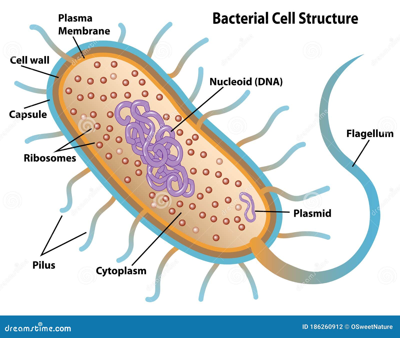
Before plunk into the mark summons, it is essential to understand the introductory construction of a bacterial cell. Bacteria are prokaryotic organisms, meaning they lack a true nucleus and other membrane bound organelles. The key components of a bacterial cell include:
- The cell wall, which provides structural support and security.
- The plasma membrane, which regulates the movement of substances in and out of the cell.
- The cytoplasm, where several metabolous processes occur.
- The nucleoid, which contains the genetic material (DNA).
- Ribosomes, which are the sites of protein synthesis.
- Flagella and pili, which are involved in motility and attachment, severally.
Techniques for Labeling A Bacterial Cell
Labeling a bacterial cell involves using assorted techniques to mark specific components. These techniques can be generally categorized into fluorescent pronounce, immunolabeling, and genetic labeling. Each method has its advantages and is chosen based on the specific component being studied.
Fluorescent Labeling
Fluorescent judge is a widely used technique that involves attach fluorescent dyes to specific cellular components. These dyes emit light when stir by a specific wavelength, allowing researchers to visualize the mark components under a fluorescence microscope. Common fluorescent dyes include:
- Fluorescein isothiocyanate (FITC)
- Rhodamine
- Cy3 and Cy5
Fluorescent labeling is particularly utile for examine the locating and dynamics of proteins, nucleic acids, and other biomolecules within the bacterial cell.
Immunolabeling
Immunolabeling, also known as immunofluorescence, involves using antibodies to specifically bind to target molecules within the cell. The antibodies are then labeled with fluorescent dyes or enzymes that create a noticeable signal. This technique is extremely specific and sensitive, making it idealistic for canvass the distribution and interactions of proteins.
The procedure of immunolabeling typically involves the following steps:
- Fixation: The bacterial cells are fixed to preserve their structure and prevent degradation.
- Permeabilization: The cell membrane is permeabilized to permit antibodies to enter the cell.
- Blocking: Non specific adhere sites are blocked to cut background noise.
- Primary Antibody Incubation: The primary antibody, specific to the target molecule, is hatch with the cells.
- Secondary Antibody Incubation: A secondary antibody, mark with a fluorescent dye or enzyme, is incubated with the cells to bind to the master antibody.
- Detection: The labeled cells are visualized under a fluorescence microscope.
Note: Proper regression and permeabilization are all-important for successful immunolabeling. Inadequate fixation can lead to loss of cellular construction, while insufficient permeabilization can prevent antibodies from access intracellular targets.
Genetic Labeling
Genetic pronounce involves insert genes that encode fluorescent proteins into the bacterial genome. These proteins, such as green fluorescent protein (GFP), can be fused to target proteins, allowing their location and dynamics to be study in living cells. Genetic label is especially utile for long term studies and for chase the demeanor of proteins in existent time.
The operation of genetic mark typically involves the follow steps:
- Construction of a plasmid: A plasmid containing the gene for the fluorescent protein is constructed.
- Transformation: The plasmid is insert into the bacterial cells through transformation.
- Selection: Bacteria that have successfully taken up the plasmid are selected using antibiotic impedance markers.
- Expression: The fluorescent protein is express in the bacterial cells, allowing the labeled component to be visualized.
Note: Genetic labeling requires measured design of the plasmid to guarantee proper reflexion and location of the fluorescent protein. Additionally, the use of appropriate promoters and regulatory elements is indispensable for controlling the expression of the fluorescent protein.
Applications of Labeling A Bacterial Cell
Labeling a bacterial cell has numerous applications in research and industry. Some of the key applications include:
Studying Protein Localization and Dynamics
By labeling specific proteins, researchers can study their fix within the cell and track their movements over time. This info is crucial for understanding the functions and interactions of proteins in several cellular processes.
Investigating Cellular Structures
Labeling techniques can be used to visualize and study the construction and organization of cellular components, such as the cell wall, plasma membrane, and nucleoid. This info is essential for understanding the architecture and function of the bacterial cell.
Diagnostic and Therapeutic Applications
Labeling a bacterial cell can also have diagnostic and therapeutic applications. for case, fluorescently labeled antibodies can be used to detect specific bacterial pathogens in clinical samples. Additionally, transmissible labeling can be used to germinate bacterial strains that create alterative proteins or function as live vaccines.
Challenges and Limitations
While label a bacterial cell is a potent creature, it also presents various challenges and limitations. Some of the key challenges include:
Specificity and Sensitivity
Ensuring the specificity and sensibility of mark techniques is crucial for accurate and reliable results. Non specific tie and background noise can lead to false plus results, while low sensibility can effect in false negative results.
Cell Viability
Some labeling techniques, such as fixation and permeabilization, can affect the viability of bacterial cells. This can limit the use of these techniques for studying living cells and dynamic processes.
Technical Complexity
Labeling techniques can be technically complex and require specialized equipment and expertise. This can be a barrier for researchers who are new to the battleground or have bound resources.
Future Directions
Despite the challenges, the field of bacterial cell labeling continues to evolve, with new techniques and technologies being developed. Some of the futurity directions in this battlefield include:
Advanced Imaging Techniques
Advances in picture technologies, such as super resolve microscopy and live cell imaging, are enable researchers to study bacterial cells with unprecedented detail and declaration. These techniques allow for the visualization of subcellular structures and dynamic processes in real time.
Multiplex Labeling
Multiplex mark involves the simultaneous judge of multiple targets within the same cell. This technique allows researchers to study the interactions and co fix of different proteins and biomolecules, providing a more comprehensive realize of cellular processes.
Single Cell Analysis
Single cell analysis involves studying individual bacterial cells to see the heterogeneity and variability within a universe. This approach is particularly utile for studying bacterial communities and their interactions with the environment.
Labeling a bacterial cell is a fundamental technique in microbiology that provides valuable insights into the structure and function of these microorganisms. By using various label techniques, researchers can study the fix and dynamics of proteins, investigate cellular structures, and develop diagnostic and therapeutic applications. While there are challenges and limitations to this approach, ongoing advancements in imaging technologies and labeling methods are pave the way for new discoveries and applications in the field of microbiology.
Related Terms:
- typical bacterium label its parts
- bacterial cell labeled year 9
- label bacteria cell worksheet
- tag bacteria cell diagram
- distinctive bacteria labeled
- bacteria drawing with labels