Understanding the intricacies of the human brain has always been a capture and challenge try. Advances in medical engineering have made it potential to explore the brain's construction and function in unprecedented detail. One of the most significant developments in this battleground is the variety of types of brain scans available today. These scans render priceless insights into neurological conditions, brain injuries, and cognitive functions. This post will delve into the different types of brain scans, their applications, and how they contribute to modern medicine.
Understanding the Basics of Brain Scans
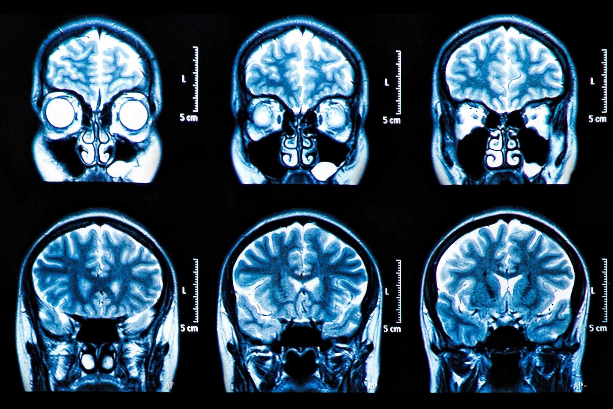
Brain scans are symptomatic tools that grant aesculapian professionals to visualize the brain's structure and action. They are crucial for diagnosing and monitor several neurological conditions, planning surgical procedures, and conducting research. The main types of brain scans include Magnetic Resonance Imaging (MRI), Computed Tomography (CT), Positron Emission Tomography (PET), and Functional MRI (fMRI). Each of these techniques offers unparalleled advantages and is used in different clinical and research settings.
Magnetic Resonance Imaging (MRI)
MRI is one of the most commonly used types of brain scans. It uses powerful magnets and radio waves to make detailed images of the brain's structure. MRI scans are particularly utilitarian for detecting tumors, aneurysms, and other abnormalities. They cater eminent resolve images that can reveal even the smallest details of brain tissue.
There are various subtypes of MRI, including:
- Structural MRI: Provides detailed images of the brain's anatomy.
- Functional MRI (fMRI): Measures brain action by observe changes in blood flow.
- Diffusion Tensor Imaging (DTI): Maps the brain's white matter tracts.
Note: MRI scans are non invasive and do not use ionizing radiation, do them a safer option for repeated imaging.
Computed Tomography (CT)
CT scans, also known as CAT scans, use X rays to make cross sectioned images of the brain. They are particularly useful for detecting acute conditions such as strokes, hemorrhages, and traumatic brain injuries. CT scans are often the first choice in emergency situations due to their speed and availability.
CT scans are less detailed than MRI scans but are faster and more widely available. They are much used to complement MRI scans in diagnosing complex neurologic conditions.
Note: CT scans use ionise radiation, so they are typically used when the benefits outweigh the risks, such as in emergency situations.
Positron Emission Tomography (PET)
PET scans use a radioactive tracer to fancy metabolous processes in the brain. They are specially utile for find cancer, assessing brain office, and studying neurological disorders such as Alzheimer's disease and Parkinson's disease. PET scans ply info about the brain's biochemical action, get them invaluable for research and diagnosis.
PET scans are often unite with CT or MRI scans to provide both structural and functional info. This combination allows for a more comprehensive understanding of the brain's condition.
Note: PET scans involve the use of radioactive tracers, so they are typically used when the benefits outweigh the risks, such as in name crab or assessing brain function.
Functional Magnetic Resonance Imaging (fMRI)
fMRI is a specialise type of MRI that measures brain action by find changes in blood flow. It is specially utile for studying cognitive functions, such as language, memory, and motor control. fMRI scans render real time info about brain action, making them priceless for research and clinical applications.
fMRI scans are non invasive and do not use ionize radiation, making them a safer option for recur project. They are often used in junction with other types of brain scans to provide a more comprehensive realise of the brain's structure and use.
Note: fMRI scans are highly sensitive to movement, so patients must remain still during the procedure.
Other Types of Brain Scans
besides the principal types of brain scans, there are several other imaging techniques that are used in specific clinical and research settings. These include:
- Electroencephalography (EEG): Measures electric activity in the brain using electrodes placed on the scalp.
- Magnetoencephalography (MEG): Measures magnetic fields produced by electrical activity in the brain.
- Single Photon Emission Computed Tomography (SPECT): Uses gamma rays to create 3D images of brain function.
Each of these techniques offers singular advantages and is used in different clinical and inquiry settings. for case, EEG is oft used to diagnose epilepsy, while MEG is used to study brain office in real time.
Applications of Brain Scans
The applications of types of brain scans are vast and varied. They are used in a wide range of clinical and research settings, including:
- Diagnosis of Neurological Conditions: Brain scans are essential for diagnosing conditions such as tumors, strokes, and traumatic brain injuries.
- Monitoring Treatment Progress: Brain scans can be used to monitor the progress of treatment for conditions such as crab and neurological disorders.
- Research: Brain scans are invaluable for research into brain function, cognitive processes, and neurologic disorders.
- Surgical Planning: Brain scans cater detail images that are essential for plan surgical procedures, such as tumor removal or deep brain stimulation.
Brain scans are also used in forensic settings to assess brain damage in cases of suspected abuse or neglect. They can ply valuable grounds in legal proceedings and facilitate to ensure that justice is serve.
Choosing the Right Type of Brain Scan
Choosing the right type of brain scan depends on various factors, include the clinical interrogative, the patient's stipulation, and the accessibility of the imaging technique. for instance, MRI scans are frequently the first choice for detail imaging of brain construction, while CT scans are used in emergency situations due to their speed and accessibility. PET scans are used to assess brain function and detect crab, while fMRI scans are used to study cognitive processes.
In some cases, a combination of types of brain scans may be used to ply a more comprehensive understand of the brain's precondition. for illustration, a PET scan may be combined with a CT or MRI scan to provide both structural and functional information.
Note: The choice of brain scan should be made in reference with a aesculapian professional who can assess the patient's precondition and determine the most appropriate figure technique.
Future Directions in Brain Imaging
The field of brain envision is rapidly evolve, with new technologies and techniques being acquire all the time. Some of the most promising areas of inquiry include:
- Advanced MRI Techniques: New MRI techniques, such as dissemination tensor visualise (DTI) and functional connectivity MRI (fcMRI), are providing unprecedented insights into brain structure and role.
- Hybrid Imaging: Combining different types of brain scans, such as PET and MRI, is render a more comprehensive understanding of the brain's condition.
- Artificial Intelligence: AI is being used to analyze brain images and identify patterns that are not visible to the human eye. This is leading to new insights into brain function and the development of new symptomatic tools.
As these technologies proceed to develop, they will provide even more detailed and accurate info about the brain's construction and function. This will lead to new diagnostic tools, treatment options, and a deeper understanding of the brain.
Note: The futurity of brain image is bright, with new technologies and techniques being developed all the time. These advances will take to new insights into brain use and the development of new diagnostic tools and treatment options.
Comparing Types of Brain Scans
To wagerer understand the differences between the various types of brain scans, let's compare them in a table:
| Type of Brain Scan | Principle | Applications | Advantages | Disadvantages |
|---|---|---|---|---|
| MRI | Magnetic fields and radio waves | Detecting tumors, aneurysms, and other abnormalities | High resolution images, non incursive | Longer scan time, not worthy for patients with metallic implants |
| CT | X rays | Detecting strokes, hemorrhages, and traumatic brain injuries | Fast, widely available | Lower resolution, uses ionize radiation |
| PET | Radioactive tracers | Detecting cancer, assessing brain purpose | Provides biochemical info | Uses radioactive tracers, lower resolution |
| fMRI | Changes in blood flow | Studying cognitive functions | Non incursive, existent time info | Highly sensible to movement |
This table provides a quick reference for the different types of brain scans, their principles, applications, advantages, and disadvantages. It can be a utile puppet for medical professionals and researchers when prefer the most appropriate project technique for a particular clinical or research specify.
Note: The choice of brain scan should be made in consultation with a aesculapian professional who can assess the patient's condition and find the most seize fancy technique.
Final Thoughts
to summarize, the variety of types of brain scans available today provides priceless insights into the brain s construction and part. From MRI and CT scans to PET and fMRI, each technique offers unique advantages and is used in different clinical and enquiry settings. As technology continues to approach, these imaging techniques will become even more precise and instructive, leading to new symptomatic tools, treatment options, and a deeper realize of the brain. The futurity of brain visualise is bright, and the possibilities are endless.
Related Terms:
- how do brain scans act
- types of brain scan tests
- let a brain scan
- brain scan images
- how to scan brain action
- brain scan that shows action